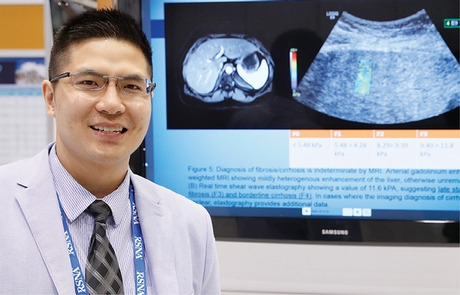
Shear Wave Elastography Via Ultrasound Offers a Painless Liver 'Biopsy'
Thursday, Dec. 01, 2016
When it comes to assessing and staging fibrosis in chronic liver disease, histopathology is still considered to be the gold standard. But what if ultrasound (US) could do the job just as reliably without subjecting the patient to a painful and inconvenient biopsy?
The Lahey Clinic in Burlington, Mass., is using a US technique, shear wave elastography, to supplement, and in many cases replace, liver biopsy. The clinic uses the technique for about 50 cases per month.
In a poster presentation on Wednesday, radiology resident Pauley Chea, MD, said most healthcare facilities should be able to use shear wave elastography with their existing US equipment, with at most a software upgrade.

Pauley Chea, MD
"Switching over can happen fairly quickly — it's just a matter of deciding whether it's what a facility needs," he said.
Chronic liver disease, including alcoholic liver disease, fatty liver disease, hepatitis, cirrhosis, cholangitis and hemochromatosis, is responsible for 1.2 percent of deaths per year in the U.S., and cirrhosis alone accounts for 35,000 deaths.
Early detection and staging of fibrosis and inflammation is key in determining prognosis and treatment outcomes, and fibrosis can be reversed if detected and treated early.
The most common causes of fibrosis are hepatitis B and C, alcoholic liver disease and non-alcoholic fatty liver disease. Fibrosis is most commonly confirmed with a biopsy. But biopsies pose their own risks — most commonly bleeding — and may yield inconsistent specimens. The biopsied site may not represent the liver's overall condition if the liver isn't homogeneous. And biopsies may be contraindicated for some patients.
Shear waves are generated in tissues when a directional force (such as US) is applied and causes deformation. Shear waves produce micrometer-level tissue displacement that can be detected by the US probe. Shear waves will propagate faster in stiffer tissues, such as a cirrhotic liver, and therefore increased propagation indicates fibrosis.
Quantified values of liver stiffness are obtained in kilopascals (kPA), a unit of pressure measurement that can be converted to the METAVIR scale (F0 to F4) that is used to grade histopathology specimens. When performed with either complete or limited abdominal US, shear-wave elastography is non-invasive and quick, samples a larger area than a biopsy, is highly reproducible, has limited dependency on the skill of the operator, and produces quantitative data.
"The beauty is that it's simple to read," Dr. Chea said. "Essentially, you place the probe in a particular position and press a button." The machine averages 10 data points to get an overall measure of stiffness.
Shear-wave elastography may be indicated when liver fibrosis is suspected, as well as hepatitis, non-alcoholic steatohepatitis, primary biliary cholangitis or other liver disease. Because fat and fluid interfere with shear wave propagation, elastography may be less accurate in obese patients, and in the presence of ascites, steatosis, inflammation, acute hepatitis, and cholestasis.
Dr. Chea predicted that the "painless biopsy" will be particularly useful to measure the effectiveness of treatment over time. Rather than requiring a biopsy every three years, hepatologists can order the scans to monitor response non-invasively, and can also avoid exposing the patient to excess radiation via repeated CT scans, or incurring the greater expense of repeat MRI studies.
The technique also might be used to evaluate blood vessels, focal liver lesions and lung fibrosis.


 Home
Home Program
 Exhibitors
 My Meeting
 Virtual
 Digital Posters
 Case of Day

